These are organs that filter out waste products from the blood to excrete in the urine.
- Cortex – tissue that processes blood to make urine
- Medulla – drains the urine from the cortex into small tubes
- Renal pelvis - collects the urine before it drains out of the kidneys (the calyces are branches of the renal pelvis)
Ureters
Pipe that drains the urine from the renal pelvis and takes it from the kidney to the bladder
- The part where the renal pelvis joins the ureter is called the pelvi-ureteric junction (PUJ)
- The part where the ureter drains into the bladder is called the vesico-ureteric junction (VUJ)
Bladder
The organ that fills and stores urine until it is convenient to empty
Urethra
Pipe that drains the bladder when urine is passed
.png)
What are CAKUT?
Congenital anomalies of the kidneys and urinary tract (CAKUT) are a number of different abnormalities in the way that the kidneys are made or in the way that they drain urine into or out of the bladder. These are often detected during pregnancy and can impact how your baby is managed after they are born.
The more common conditions include:
- Renal pelvis dilatation
- Multicystic dysplastic kidney
- Absent kidney
- Ectopic or horseshoe kidney
- Duplex kidney with or without a ureterocoele
What is renal pelvis dilatation?

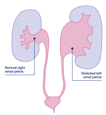
Renal pelvis dilatation (or hydronephrosis) is a widening of the renal pelvis and is a common finding on ultrasound scans (USS) performed during pregnancy. Often it is temporary and not associated with any problems in the kidney or ureter. In this situation, there is no risk for your child’s future health.
However, in a small proportion of cases, renal pelvis dilatation can be a sign of problems involving the kidneys, ureters, bladder, or the urethra. The most common conditions involve either a blockage in the pipes that drain the urine or refluxing of urine back up the pipes towards the kidneys (Vesicourethral Reflex (VUR).
Causes of renal pelvis dilatation:
- PUJ Obstruction: a partial obstruction at the junction between the renal pelvis and the ureter which can restrict the flow of urine to the bladder. If the obstruction is severe, it can stop the kidney from working normally
- VUJ Obstruction: a partial obstruction at the junction between the ureter and the bladder which can restrict the flow or urine into the bladder. If the obstruction is severe, it can stop the kidney from working normally
- Vesico-ureteric reflux (VUR): normally urine travels only one way down the ureter - from the kidney to the bladder. At the point where the ureter enters the bladder there is a valve that normally closes when the bladder contracts in preparation for passing urine. If the closure is not complete then urine can travel back up to the kidney. This is called reflux. Reflux can cause dilation of the ureter, the renal pelvis and if severe can stop the kidney from working normally or increase the risk of baby contracting a urine infection.
- Posterior urethral valves (PUV): a condition that only occurs in boys where abnormal tissue in the urethra prevents urine passing from the bladder. This can cause pressure to build up in the bladder and urine to reflux up one or usually both ureters and cause dilatation of the renal pelvis and can stop the kidneys from working normally. This can be a serious condition that requires immediate treatment after birth.
Other conditions that can cause renal pelvis dilatation are also possible and your doctor will discuss these conditions with you in more detail if required.
What are the other congenital anomalies of the kidney and urinary tract?
The most common forms of CAKUT include:
.png)
- Multicystic Dysplastic Kidney: a condition where the normal functioning kidney tissue has been replaced by fluid filled cysts. This usually happens very early in the development of that kidney and that kidney will never work. Usually the kidney will shrink and be completely absorbed by the body, sometimes even before baby is born. It is rare, but sometimes, if the kidney does not shrink and is very large, an operation is required to remove that kidney. Although most people are born with two normal functioning kidneys, having only one normal working kidney should have very little effect on your baby’s life. An appointment with the kidney specialists in Queensland Children’s Hospital will be organised after baby is born.
- Absent Kidney: Around 1 in every 750 people will only have one kidney. Many are born with only one kidney because either the kidney was never made on that side (renal agenesis) or because there was a multicystic dysplastic kidney that shrunk and was absorbed before baby was born. Others are born with a multicystic dysplastic kidney that shrinks and is absorbed after baby is born. As long as the other kidney is normal this should have very little effect on your baby’s life. An appointment with the kidney specialists in Queensland Children’s Hospital will be organised after baby is born.
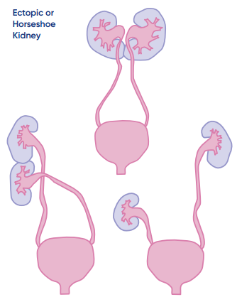
- Ectopic Kidney or Horseshoe Kidney: An ectopic kidney is one that is not in the normal place. Ectopic kidneys are
 commonly found lower in the pelvis (pelvic kidney), but can also be found higher up under the diaphragm and sometimes one kidney can be found across the other side stuck to the normally placed kidney (crossed-fused ectopic kidney). When both kidneys are placed closer to the middle and stuck together they are called a horseshoe kidney. As long as both kidneys are working normally and there is no blockage to the flow of urine, having an ectopic or horseshoe kidney should have very little effect on your baby’s life. However, because there is an increased risk of problems with either kidney, before you leave hospital we will arrange for an ultrasound scan to be performed when your baby is a few weeks old.
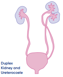
- Duplex Kidney and Ureterocoele: A duplex kidney occurs when the kidney develops in two separate parts and there are often two collecting systems and two ureters. The two ureters may travel completely separately into the bladder (like the picture) or come together and join before entering the bladder. Having a duplex kidney affects about 1 in a 100 people and in half of the cases both kidneys are duplex. Having a duplex kidney is often associated with one of the ureters entering the bladder in the wrong place (ectopic ureter) and this can also lead to a bubble forming inside the bladder at the point that the ureter enters (ureterocoele). Although having a duplex kidney does not cause any problems in itself it is associated with a number of problems including a blockage either where the ureter comes out of the kidney or if there is a
 ureterocoele this can block the passage of urine into the bladder. If there is a blockage this may mean that your baby will require surgery to relieve the obstruction to the flow of urine. Duplex kidneys are also prone to vesicoureteric reflux (see previous page under “causes of renal pelvis dilatation”). Because of this increased risk of problems with either kidney, before you leave hospital we will arrange for an ultrasound scan to be performed when your baby is a few weeks old.
What happens if a congenital anomaly of the kidney and urinary tract is discovered?
Usually, the first step is an ultrasound scan of the kidneys (renal USS) after your baby is born. For most babies, the best time to do this is at around 4 weeks of age. For some babies the ultrasound scan will need to be performed before your baby is discharged home. For some anomalies, an ultrasound may not be required and an appointment with one of the kidney specialists in Queensland Children’s Hospital will be organised for when your baby is around 3 months of age. The team caring for your baby will explain the best plan for your baby after they are born.
If your baby requires an ultrasound scan it will either be here at Mater Hospital before your baby goes home, or at Queensland Children’s Hospital in a few weeks’ time. If the scan is done in Mater Hospital, the team caring for your baby will explain the results and the plan for treatment and follow up before baby is discharged home. If your baby has the scan after going home, you will receive a follow up from Mater or Queensland Children’s team with the results of ultrasound scan, as well as an explanation of any further tests that may be required and any other follow up that might be organised.
How to recognise if your child has a UTI
It can be hard to know when your child has a UTI. If your child has a fever it is advisable to see your general practitioner (GP) so your child can be reviewed and their urine tested.
Some signs your child may have a UTI include:
- fever
- poor feeding
- being irritable or more tired and sleepy than usual
- sore stomach.
If your child does have a UTI then further tests may be needed and you can discuss this with your GP. Appropriate early detection and management of conditions involving the kidney/bladder systems may prevent or reduce future injury or abnormal kidney development for your child.
Prophylactic antibiotics
In some conditions a low dose of an antibiotic medicine, given once a day (best given at night), may be recommended. This is to reduce the chance of urinary tract infection (UTI) occurring. Many conditions, including mild to moderate renal pelvis dilatation have not been shown to benefit from having prophylactic antibiotics.
© 2023 Mater Misericordiae Ltd. ACN 096 708 922